Home

difficile Tipicamente imparare fetal pole picture umorismo diminuire lucidatura

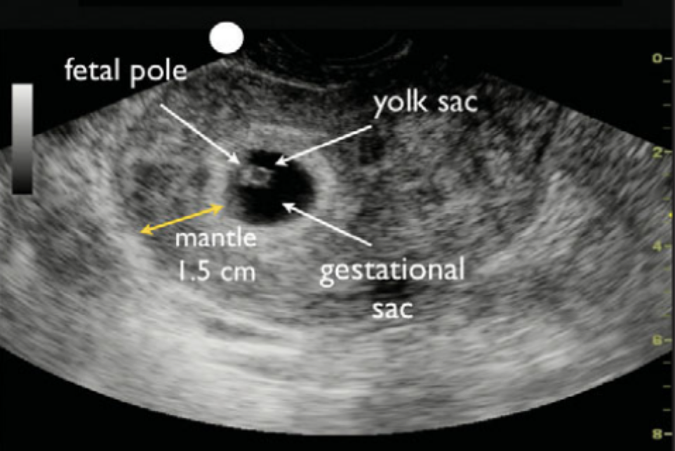
Yolk sac and fetal pole not seen meaning in hindi | Empty gestational sac -  YouTube
Yolk sac and fetal pole not seen meaning in hindi | Empty gestational sac - YouTube

Yolk sac but no fetal pole - June 2021 Babies | Forums | What to Expect

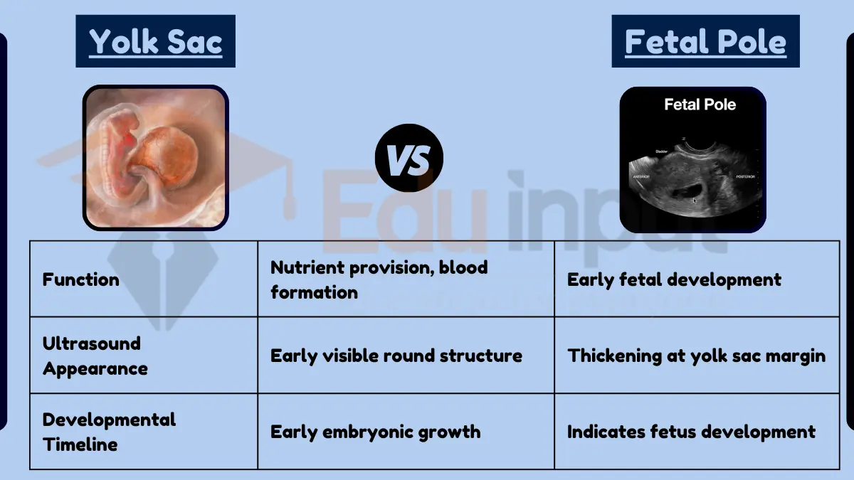
What is the Difference Between Yolk Sac and Fetal Pole | Compare the  Difference Between Similar Terms
What is the Difference Between Yolk Sac and Fetal Pole | Compare the Difference Between Similar Terms
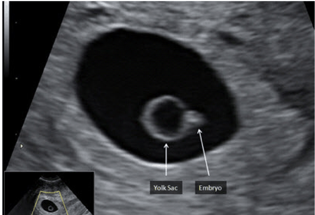
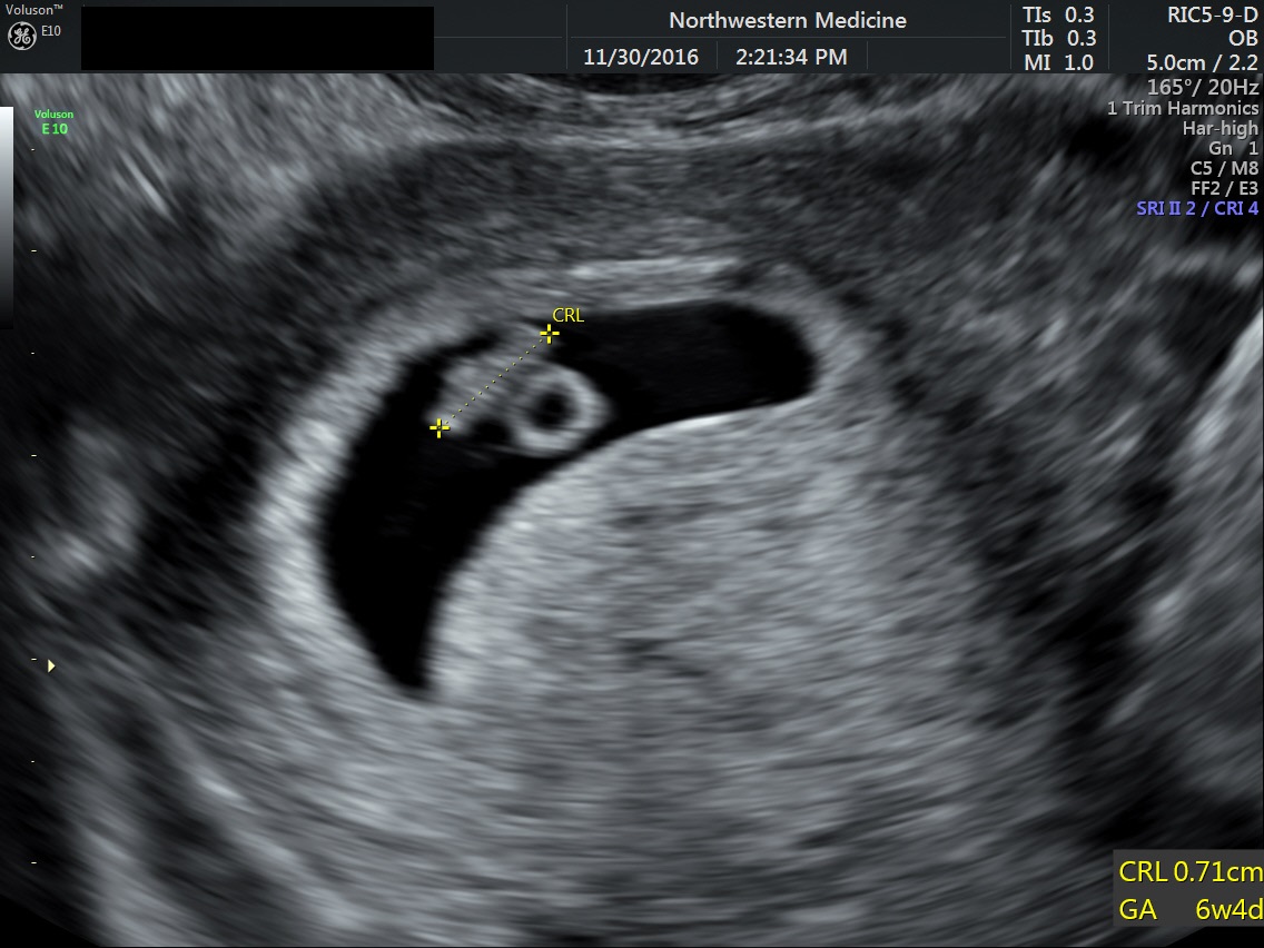
POCUS 101 on X: "(6/n) Fetal Pole appears around 5.5-6 weeks (transvaginal  US) 👉🔗https://t.co/wBsqQSYLfk https://t.co/VRdGbp1pIW" / X
POCUS 101 on X: "(6/n) Fetal Pole appears around 5.5-6 weeks (transvaginal US) 👉🔗https://t.co/wBsqQSYLfk https://t.co/VRdGbp1pIW" / X

Definitive IUP - Fetal Pole - YouTube

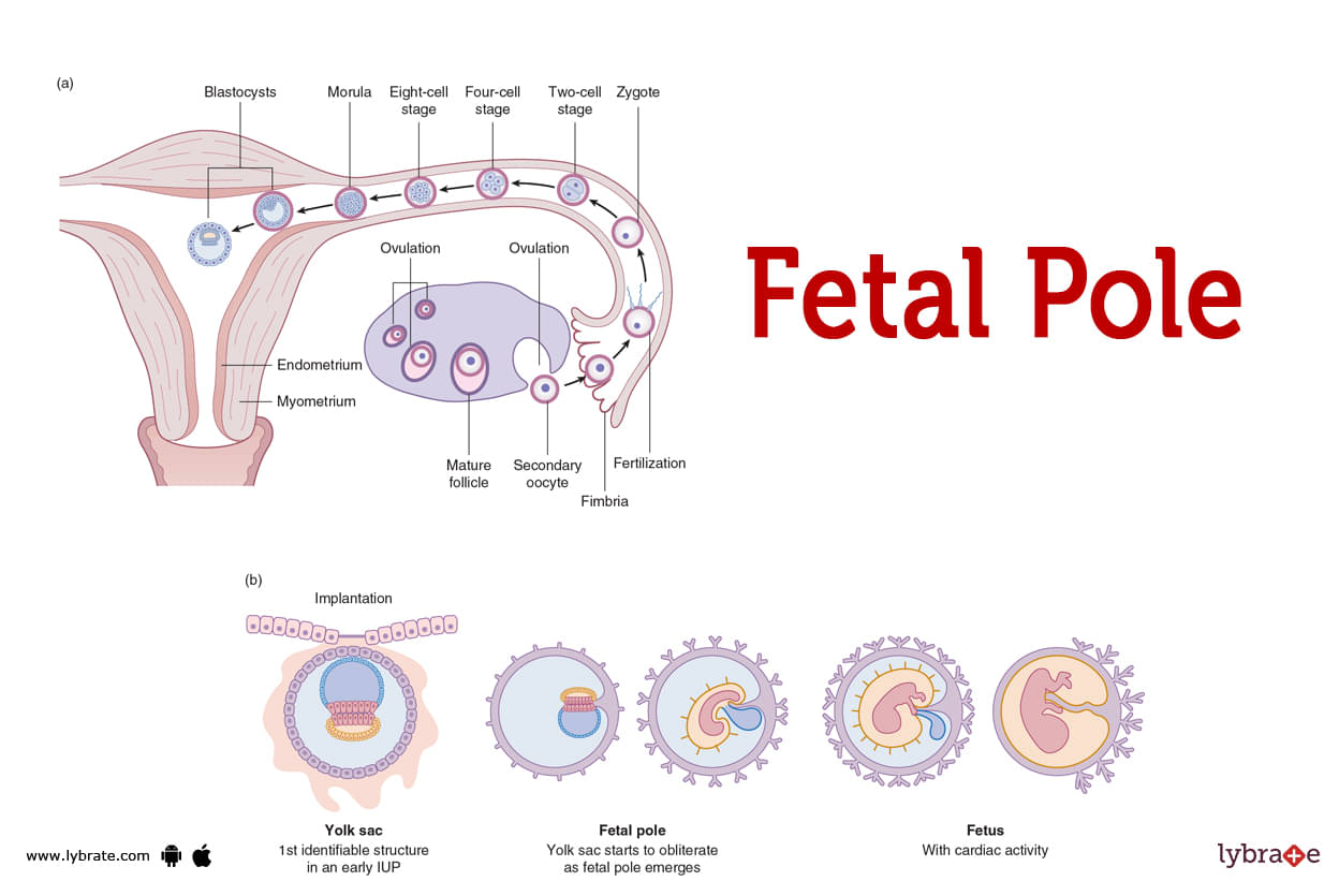
Fetal Pole (Human Anatomy): Image, Functions, Diseases and Treatments

Pregnancy: Week 6 | By London Pregnancy Clinic | Medium

Diagnostic Ultrasound in the First Trimester of Pregnancy | GLOWM

Difference Between Yolk Sac and Fetal Pole

Fetal Pole (Human Anatomy): Image, Functions, Diseases and Treatments

Gestational Sac No Fetal Pole | TikTok

Abnormal Ultrasound in Early Pregnancy

The embryo is first visible as a fetal pole adjacent to the yolk sac... |  Download Scientific Diagram
The embryo is first visible as a fetal pole adjacent to the yolk sac... | Download Scientific Diagram

ALiEM Cards
ALiEM Cards

Diagnostic Ultrasound in the First Trimester of Pregnancy | GLOWM

OBSTETRIC (First Trimester) | Department of Emergency Medicine | Saint John

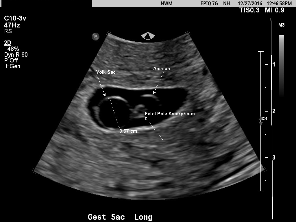
A) Normal fetal pole (yellow arrow) and YS (blue arrow) in a patient... |  Download Scientific Diagram
A) Normal fetal pole (yellow arrow) and YS (blue arrow) in a patient... | Download Scientific Diagram
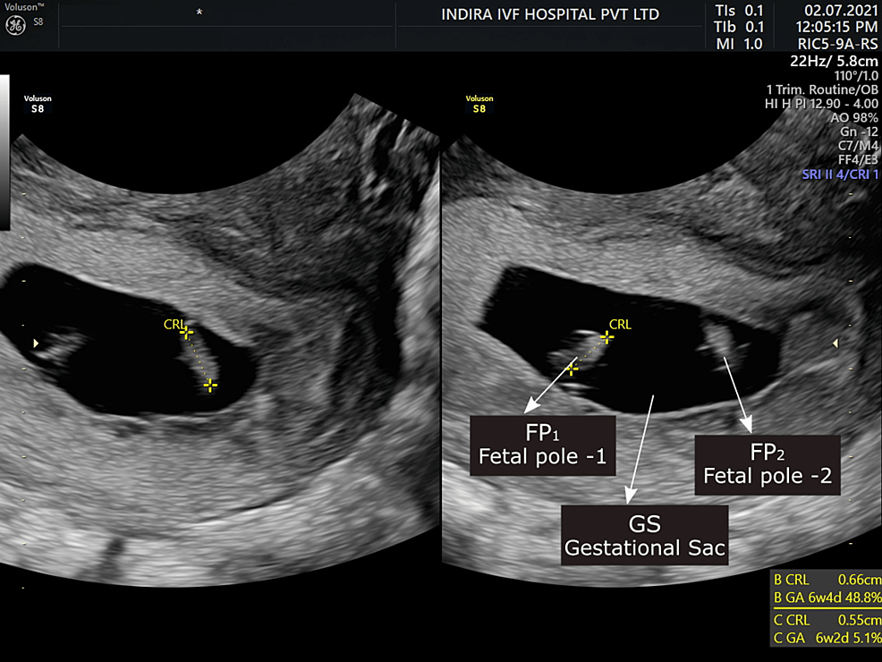
Cureus | Successful Management of Triplet Heterotopic Pregnancy  (Interstitial) With an Intrauterine Monochorionic Diamniotic Twin Pregnancy  Through Laparoscopy: A Case Report | Media
Cureus | Successful Management of Triplet Heterotopic Pregnancy (Interstitial) With an Intrauterine Monochorionic Diamniotic Twin Pregnancy Through Laparoscopy: A Case Report | Media

Case of the Month — Ectopic Pregnancy — BROWN EMERGENCY MEDICINE

Fetal sex prediction measuring yolk sac size and yolk sac–fetal pole  distance in the first trimester via ultrasound screening | Journal of  Ultrasound
Fetal sex prediction measuring yolk sac size and yolk sac–fetal pole distance in the first trimester via ultrasound screening | Journal of Ultrasound

A wide fetal pole with bifid appearance and two yolk sacs in a unique... |  Download Scientific Diagram
A wide fetal pole with bifid appearance and two yolk sacs in a unique... | Download Scientific Diagram

Fetal pole of intrauterine pregnancy, Twin A. | Download Scientific Diagram
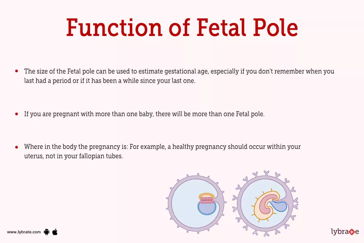

Fetal Pole and Early Pregnancy Ultrasound

Gravid uterus with a fetal pole corresponding to about 7 weeks | Download  Scientific Diagram
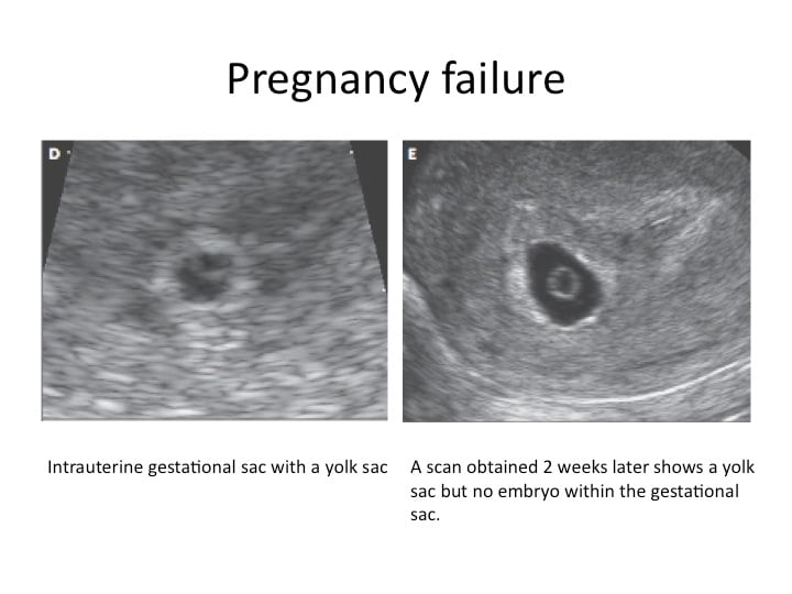
Gravid uterus with a fetal pole corresponding to about 7 weeks | Download Scientific Diagram
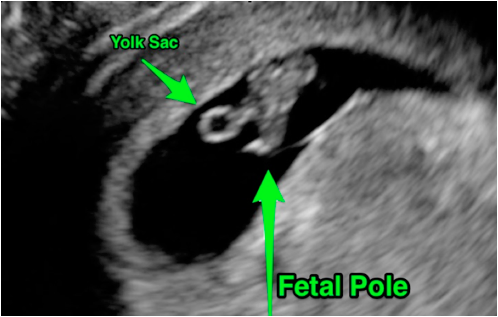
No fetal pole at 6 weeks - any stories? | Mumsnet

Figure 1. | Meningomyelocele: Early Detection Using 3-Dimensional  Ultrasound Imaging in the Family Medicine Center | American Board of Family  Medicine
Figure 1. | Meningomyelocele: Early Detection Using 3-Dimensional Ultrasound Imaging in the Family Medicine Center | American Board of Family Medicine